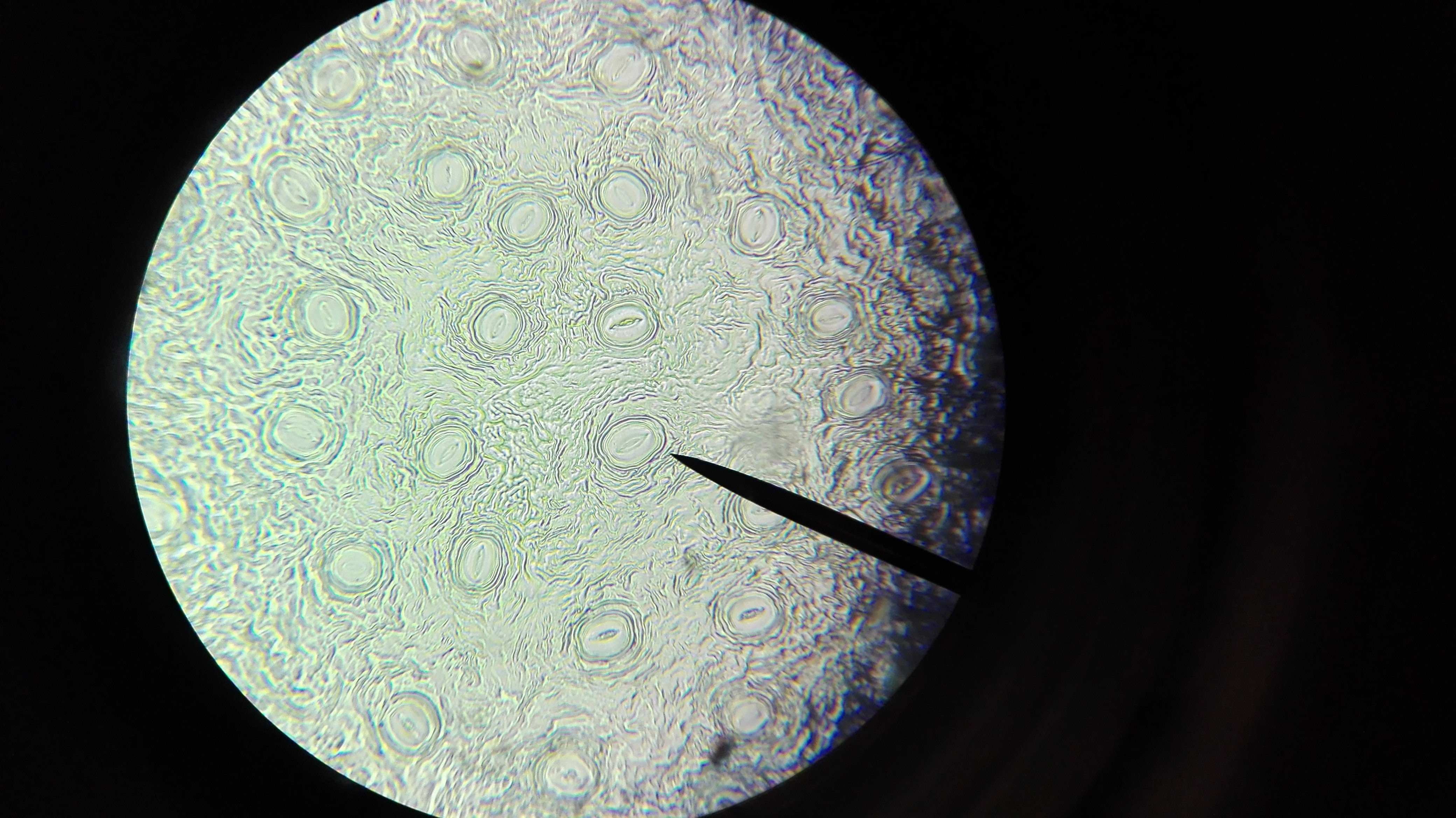
The development of microscopy has shaped cell biology since its inception. With each technical improvement, the view into the inside of the cell became sharper and new structures were discovered. The term ‘cell’ was coined by Robert Hooke as early as the 17th century. He provided the first descriptions in his work Micrographia (1665) after examining the porous structure of cork with an early version of a microscope (Fig. 1) [1]. A few years later, Antonie van Leeuwenhoek used high-resolution single lenses he had made himself to observe protozoa and bacteria, for which he coined the term ‘animalcules,’ thus opening up a previously unknown microbial world [2].
One would think that the cell has been thoroughly researched by now and that at least all intracellular components have been identified. But far from it: modern cryo-tomography provides insights that were previously unimaginable and has recently led to the identification of a previously unknown organelle: the hemifusom.
These topics await you:
1) From Cork to Cryo-Tomography - how Microscopes Shaped our View of Life
2) The Hemifusome - a Newly Identified Organelle
3) From the Cell to Therapy - the Potential Significance of Hemifusomes
![]()
From Cork to Cryo-Tomography - how Microscopes Shaped our View of Life
The work of Hooke and van Leeuwenhoek laid the foundation for the triumph of microscopy, which gained increasing popularity in the following decades. Advances in lens technology in the 19th century enabled scientists Matthias Schleiden and Theodor Schwann to develop the fundamentals of the cell theory. This theory states that plants and animals are both made up of cells [2]. As a result, intracellular structures increasingly became the focus of attention, such as the Golgi apparatus discovered by Camillo Golgi using silver staining [3].

The development of electron microscopy in the 20th century heralded a new era in cellular research that went far beyond the discoveries made by Hooke and van Leeuwenhoek. The first transmission electron microscope (1931) enabled unprecedented resolution of cellular structures. This was followed by phase contrast microscopy (1932) and confocal laser microscopy (1957), which allowed increasingly detailed and dynamic representations of cells and organelles [4].
Finally, in the 21st century, cryo-electron microscopy and cryo-tomography opened up access to nearly native, three-dimensional views of molecules and organelles within living systems. This development illustrates that even today, fundamental structures can still be redescribed. One example of this is the recently discovered structure of the hemifusome, which was first described in the publication ‘Hemifusomes and interacting proteolipid nanodroplets mediate multi-vesicular body formation’ (Tavakoli et al. 2025) in Nature Communications [6]. The study was conducted as part of a collaboration between the University of Virginia and the National Institutes of Health. The hemifusome is a previously unknown vesicular organelle that could only be visualised using high-resolution cryo-electron tomography [6].
The Hemifusome - a Newly Identified Organelle
Hemifusomes are vesicle complexes consisting of two unequal vesicles (Fig. 1) that are partially fused together, forming an extensive common membrane surface, which researchers refer to as the ‘hemifusion diaphragm’ (HD). Typically, a tiny proteolipid nanodroplet (PND), a drop of fat and protein substances only about 42 nanometres in size, is located at the edge of this contact point. In the high-resolution images, it looks like a small button on the membrane (Fig. 1, left). This recurring pattern of vesicle pairs, HD and PND was the key to identifying hemifusomes as a separate organelle, as it was previously believed that these structures were only randomly occurring intermediate states [6].
What is particularly exciting is that hemifusomes occur in two morphologically different variants: a direct form (Fig. 2, left) and an inverted form (Fig. 2, right). One could say they are like a Lego brick that can be attached from two sides, once with the opening facing outwards and once facing inwards. Despite these different orientations, the basic structure remains the same. Two vesicles share a contact surface and are linked to a PND [6].
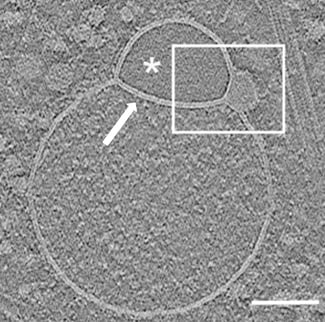
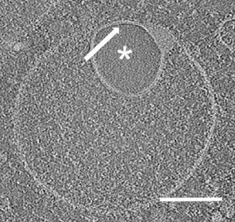
It is also surprising how frequently these structures occur. Analyses show that up to ten percent of the vesicular organelles in the cell periphery are hemifusomes. They are therefore not a rare exception, but a recurring element in the complex inner workings of the cell. The smaller of the two vesicles usually has a transparent lumen, while the larger one is filled with granular material, similar to that found in endosomes. This suggests that the smaller vesicle is filled with a very low-protein, aqueous solution, while the larger vesicle has a protein-rich content [6].
The significance of hemifusomes probably lies in the organisation of the cellular recycling system. An important component of protein sorting in the cell is the ESCRT (endosomal sorting complexes required for transport) signalling pathway. Its components promote the formation of intraluminal vesicles (ILVs) in the endosome and are thus essential for the formation of so-called multivesicular bodies (MVBs). The new observations by Tavakoli et al. suggest that hemifusomes represent an alternative, ESCRT-independent mechanism for the formation of MVBs. The close connection to proteolipid droplets could be crucial in this context: these droplets accumulate lipids and associated membrane proteins locally, thereby providing the building blocks for vesicle pinching off and stabilisation of the resulting MVBs [6].
![]()
From the Cell to Therapy - the Potential Significance of Hemifusomes
The discovery of hemifusomes is not only exciting from a cell biology perspective, but could also open up new avenues in medicine. Since they appear to play a central role in recycling and signal transmission within the cell, defective hemifusomes could be linked to various diseases. Disruptions in the endomembrane system have already been described in neurodegenerative diseases, viral infections and cancer, and hemifusomes could be a previously overlooked factor here. Future research must clarify whether the targeted manipulation of these structures reveals new therapeutic possibilities [6].
The discovery of the hemifusome shows that the cell is still far from being fully understood. Despite centuries of research, modern technology is still enabling us to discover new structures that change our understanding of life. From Hooke's view of simple cork through his microscope in the 17th century to today's cryo-tomography of cells, a line of discoveries has been drawn, and the hemifusome now fits into this picture as another surprising building block.
Sources
[1] https://www.britannica.com/technology/microscope/History-of-optical-microscopes, 28.08.2025
[2] https://education.nationalgeographic.org/resource/history-cell-discovering-cell/?utm_source=chatgpt.com, 28.08.2025
[3] https://de.wikipedia.org/wiki/Camillo_Golgi, 28.08.2025
[4] https://www.sciencelearn.org.nz/resources/1692-history-of-microscopy-timeline?utm_source=chatgpt.com, 28.08.2025
[5] https://commons.wikimedia.org/wiki/File:Hooke-microscope.png, 28.08.2025
[6] Tavakoli, A., Hu, S., Ebrahim, S. et al. Hemifusomes and interacting proteolipid nanodroplets mediate multi-vesicular body formation. Nat Commun. 16, 4609 (2025).
Preview-picture: https://pixabay.com/de/photos/mikroskop-zelle-untersuchen-1471068/
Written by Pauline Kipp


